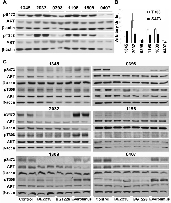
Figure 6. Phosphorylation of AKT in does not predict response to everolimus or dual kinase inhibitors.
(A) Cell lysates were prepared from xenograft cells recovered from untreated mice and subjected to Western analysis. Lysates from two mice bearing each xenograft are shown. (B) Bands were quantified by densitometry and the mean ± SE of the ratio of phosphorylated to total AKT is shown. (C) Xenograft cells were cultured for 6 hours in the presence of 2 μM everolimus, 0.2 μM BEZ235 or 0.2 μM BGT226 and cell lysates prepared. Lysates were sequentially probed for phosphorylated and total AKT, and beta-actin.

