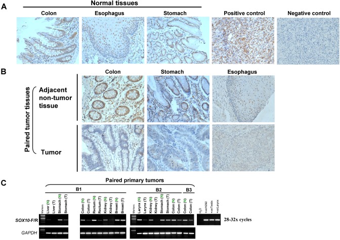
Figure 2. SOX10 expression is significantly decreased in tumor tissues.
(A) Immunohistochemistry analysis (IHC) was performed with an anti-SOX10 antibody on a human normal tissue microarray (TMA). Melanoma tissue was used as a positive control and nonimmune mouse immunoglobulin G substituted for the primary antibody as negative control (original magnification ×400). (B) SOX10 expression is significantly decreased in different tumor tissues. (C) mRNA expression levels of SOX10 in different tumor tissues (T) and their paired adjacent normal tissues (N) as determined by semiquantitative RT-PCR (28 or 32 cycles).

