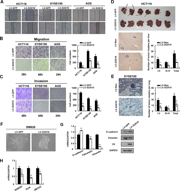
Figure 4. SOX10 suppresses tumor metastasis and EMT phenotype.
Cells were infected with LV-SOX10 or LV-GFP and subjected to wound-healing assay (A), migration assay (B) and invasion assay (C). Microscopic observations were recorded at 0 as well as 48 or 72 hours after scratching the surface of a confluent layer of cells(A). Cells that migrated (B) or invaded (C) to the lower chamber were fixed, stained, and counted using light microscopy. Values are mean ± SD for triplicate samples from a representative experiment. *: p<0.05. **: p<0.01. (D) HCT116 cells infected with LV-SOX10 or LV-GFP were inoculated into the spleen. The mice were killed and examined for the presence of hepatic metastasis 12 weeks after the intrasplenic inoculation. The upper, metastatic nodules on the surface of the liver are shown; the left, representative H&E staining of liver (original magnification, ×400); the right, the numbers of nodules were quantified and values for each group are denoted (*: p<0.05). (E) Male Balb/c nude mice were injected subcutaneously with 5×10⁶ KYSE150 cells. Representative lung tissue sections by HE-staining from each group were shown in the left (magnification ×400). The number of lung metastatic foci in each group was calculated under microscope. (F) Morphology changes of SW620 cells infected with LV-SOX10 or empty vector by phase contrast microscopy. Original magnification, ×400. (G) qRT-PCR and western blot validation of EMT biomarkers (*: p<0.05, **: p<0.01). GAPDH was used as a control. (H) Downregulation of representative stem cell markers in SOX10-infected tumor cells.

