
Figure 4. In vivo imaging of antitumor activity of CTA specific T-cells in xenograft models.
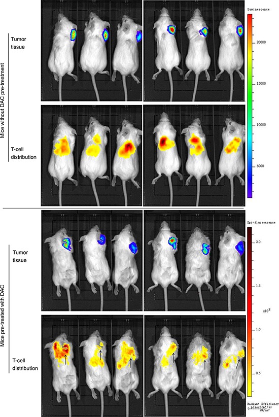
HOS cells transfected with luciferase (HOS-Luc) and SCID mice were used to establish animal models. Mice were imaged 24 hours postinjection of T-cells. Bioluminescence by HOS-Luc cells and fluorescence by DiR labeled T-cells were visualized with imaging system. In mice without DAC pre-treatment, T-cells distributed in liver, lung and scar of injection sites, while no signal was detected around the tumor tissue. In mice treated with DAC, T-cells clustered at the tumor site in addition to liver and lung. Demethylating treatment promoted the response of CTA specific T-cells to osteosarcoma tissue.