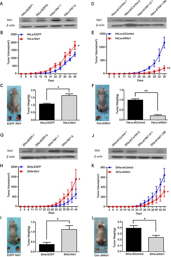
Figure 2. Msi1 enhances tumor formation by cervical cancer cells in vivo.

Western blot analysis of Msi1 protein in HeLa-EGFP and HeLa-Msi1 cells (A) HeLa-shControl and HeLa-shMsi1 cells (D) SiHa-EGFP and SiHa-Msi1 cells (G) and SiHa-shControl and SiHa-shMsi1 cells (J) β-actin served as the loading control. Tumor formation assays were performed with 6 mice per group. The tumor growth curves were calculated by monitoring that was performed every 3 days post-transplant. At 36 days post-transplant for HeLa-Msi1 cells (B) 35 days post-transplant for HeLa-shMsi1 cells (E) 44 days post-transplant for SiHa-Msi1 cells (H) and 45 days post-transplant for SiHa-shMsi1 cells (K) the xenograft tumors of mice in each group were dissociated and weighed (C, F, I and L). The data are shown as the mean± S.E.M. *P < 0.05; **P < 0.01.
