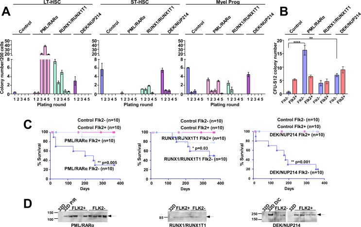
Figure 2. The effect of the AAFPs on the replating efficiency, CFU-S12 potential and the leukemogenic potential of ST- and LT-HSC.
(A) Sca1+/lin− BM cells were infected with retroviruses as indicated in reference [6]. At 12 hours post-infection, the cells were sorted for LT-HSCs (Sca1+/c-Kit+/lin−/Flk2−), ST-HSCs (Sca1+/c-Kit+/lin−/Flk2+) and myeloid progenitors (MPs) (Sca1−/c-Kit+/lin−). The sorted cells were then plated in semi-solid medium supplemented with mIL-3, mIL6 and mSCF to determine the serial replating potential. In the first plating for LT-HSC 300 cells/well, for ST-HSC 500 cells/well and for MP 3×10³ cells/well were seeded. The colonies were counted after 8–10 days, prior to each replating round, and the cell number was assessed for further replatings. For the following plating rounds 2.5×10³ cells/well were seeded. For platings with cell numbers lower than 2.5×10³ cells/well, all cells were replated. In order to keep comparable the different samples the colony counts are reported to 200 cells seeded. One representative experiment (+/−SD) is shown. Three experiments were performed in total, with similar results, and each experiment was performed in triplicate. (B) Differential effects of the AAFPs on the potential of LT- und ST-HSCs to form colonies in a CFU-S12. The cells harvested from the first plating in the semi-solid medium were inoculated into lethally irradiated recipient mice for the CFU-S12 assay. The animals were sacrificed at day 12, and the spleen colonies were counted. We show one representative experiment of at least three performed experiments (all had similar results). Each group was comprised of 3 mice. (C) Differential effects of the AAFPs on the potential of LT- und ST-HSCs to induce leukemia. The survival curves show the frequency of recipients that succumbed to disease after receiving the cells harvested from the CFU-S12 spleens. Each group contained 10 mice. (D) Expression of the AAFPs in leukemic mice inoculated with the Sca1+/c-Kit+/lin−/Flk2− (Flk2-) and in healthy mice inoculated with the Sca1+/c-Kit+/lin−/Flk2+ (Flk2+) populations, respectively, as determined by Western blot. Mock-transduced 32D cells or cells that stably expressed the respective AAFPs were used as controls.

