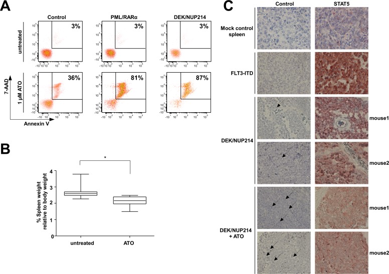
Figure 7. Responsiveness of DEK/NUP214-positive cells to ATO treatment.
(A) 7-AAD and Annexin V staining of mock-control, DEK/NUP214- and PML/RARα-positive U937 cells after treatment with 1 μM ATO for five days. Percentage of dead cells (7-AAD and Annexin V positive) is shown. (B) Frozen spleen cells isolated from DEK/NUP214-leukemic mice were inoculated into sublethally irradiated recipient mice. Five days after transplantation the mice from the treatment group received ATO for 14 days. The control group received PBS. Mice were sacrificed and spleen size was measured at the first signs of illness among the control group (n=8 mice/group) (C) Immunohistochemical staining of formalin-fixed paraffin-embedded spleen sections from mice described in 7B to determine the nuclear localization of STAT5 in untreated vs. ATO-treated group. As controls, spleen sections from mice injected with mock-transduced HSPCs or from mice with FLT3-ITD driven leukemia were used as negative and positive control, respectively.

