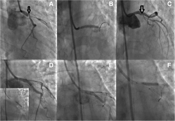
Figure 4.

PCI with TAP technique (T And Protrusion), step by step analysis. A: The true lumen was crossed and the wire was placed in the distal Cx. B: A 3.0 × 24 mm Taxus Element stent was deployed at 14 atms into the left main and proximal LCx C: Due to the proximal sealing of the dissection (arrow), there was flow improvement in the LAD D: A second Taxus Element 3.0×16mm was deployed, due to residual stenosis distally to the implanted stent of the LCx, overlapping it E,F: The procedure was concluded by placing a Taxus Element 3.0 × 16 mm stent, into the LAD, with TAP technique (T And Protrusion).
