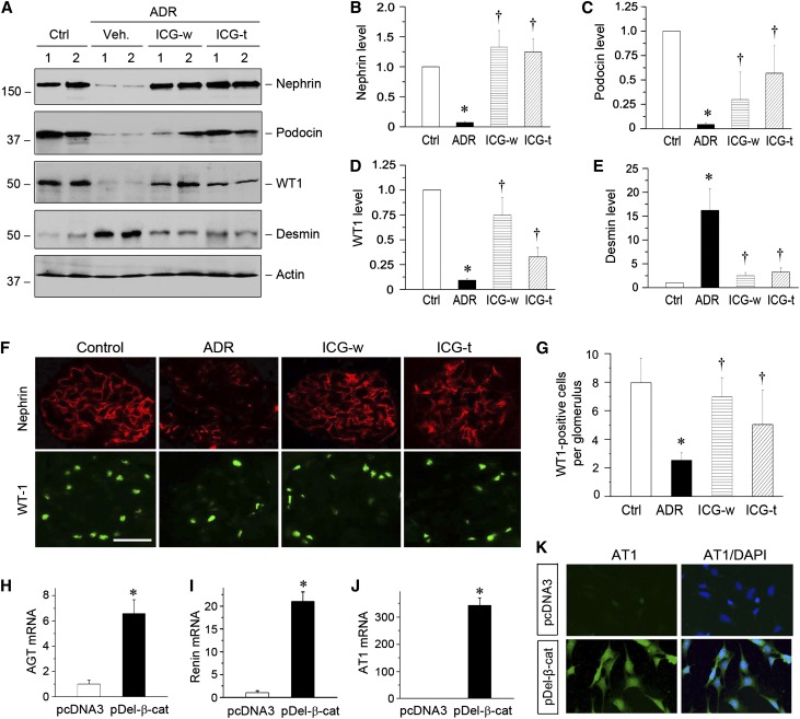
Figure 5.
ICG-001 restores podocyte integrity in ADR nephropathy. (A) ICG-001 restores podocyte-specific proteins and inhibits desmin expression. Kidney lysates were immunoblotted with specific antibodies against nephrin, podocin, WT1, desmin, and actin. Numbers 1 and 2 represent different animals in a given group as indicated. (B–E) Graphic presentations of (B) nephrin, (C) podocin, (D) WT1, and (E) desmin expressions in different groups as indicated. *P<0.05 versus normal controls; †P<0.05 versus ADR alone (n=5–6). (F) Representative micrographs showing glomerular nephrin and WT1 expression. Frozen kidney tissue sections were stained with nephrin (red) and WT1 (green). (G) Graphic presentation shows the numbers of WT1-positive podocytes per glomerular cross-section. *P<0.05 versus normal controls; †P<0.05 versus ADR alone (n=5–6). (H–J) Expression of active β-catenin induces mRNA expression of RAS genes in vitro. Mouse podocytes were transiently transfected with N-terminally truncated, FLAG-tagged, constitutively activated β-catenin expression vector (pDel-β-cat) or control pcDNA3. The mRNA expression of (H) AGT, (I) renin, and (J) AT1 was assessed by quantitative RT-PCR. *P<0.05 (n=3). (K) Expression of active β-catenin induces AT1 protein expression in cultured podocytes. Mouse podocytes after transfection were immunostained for AT1 protein (green). Cell nuclei were visualized with DAPI staining (blue). Ctrl, control; DAPI, 4′,6-diamidino-2-phenylindole; Veh, vehicle.

