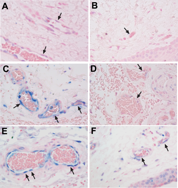
Figure 3.
Increased RAGE expression in vascular endothelial cells in human recurrent pterygium compared to corresponding conjunctiva samples. RAGE staining in pterygium (A) and conjunctiva (B) samples from case 3. RAGE staining in pterygium (C) and conjunctiva (D) samples from case 14. RAGE staining in pterygium (E) and conjunctiva (F) samples from case 15. Black arrows indicate vascular endothelial cells. The images were taken at 40X objective.

