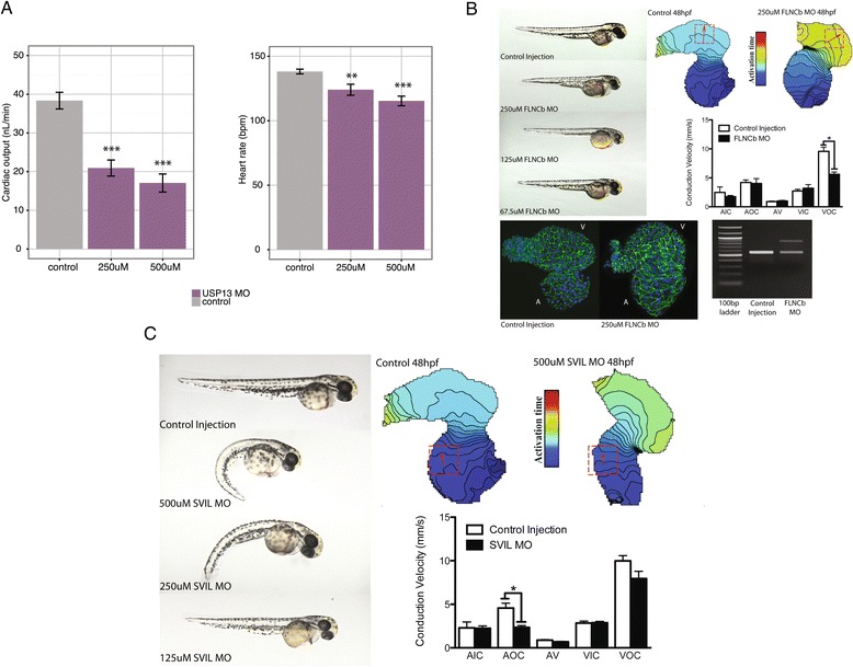
Figure 5.

OPEN prioritized genes contribute to cardiac phenotypes in zebrafish. (A) Knockdown of USP13 caused a dose-dependent decrease in cardiac output, due to both a decrease in heart rate and ventricular stroke volume. (B) Injection of a morpholino (MO) targeting a specific splicing event in FLNCb (see Materials and methods) caused apparent cardiac-specific defects. Images on the right show embryos at 48 hours post-fertilization (hpf) with decreasing injected morpholino concentration. Optical mapping confirmed a significant decrease in cardiac conduction velocity in isolated hearts following FLNCb splice inhibition (top right: isochronal maps on right, red box indicates measured region of interest, isochrones are 5 ms apart). Conduction velocity was unaltered in other regions of the heart (middle right: bar graph, regions examined were atrial inner curvature (AIC), atrial outer curvature (AOC), AV node (AV), ventricular inner curvature (VIC), and ventricular outer curvature (VOC)). Additionally, FLNCb splice inhibition resulted in increased atrial cardiomyocyte size (bottom left: beta-catenin stained in green, DAPI in blue, V and A denote ventricle and atria, respectively). RT-PCR confirmed inhibition of the predicted splicing event in FLNCb (bottom right). (C) Knockdown of SVIL causes cardiac edema as well as noticeable spinal curvature at higher morhpolino doses, with only cardiac edema notable at lower doses. Images on left again show decreasing morpholino dose at 48 hpf. Optical mapping (right) confirmed a significant decrease in atrial conduction velocity following SVIL knockdown. ***P < 0.001, **P < 0.01, *P < 0.05.
