Figure 4.

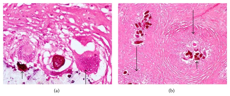
Photomicrographs showing calcified schistosome ova (short arrows), inflammatory giant cell (medium arrow), and granuloma surrounding the ova (long arrows), in the right tube.

Photomicrographs showing calcified schistosome ova (short arrows), inflammatory giant cell (medium arrow), and granuloma surrounding the ova (long arrows), in the right tube.