Figure 5.

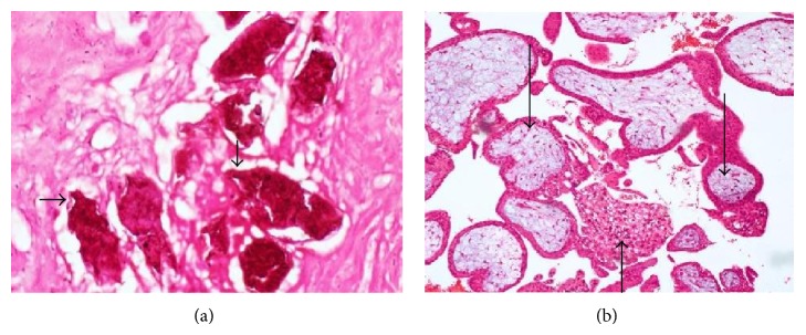
Photomicrographs showing S. haematobium ova with characteristic terminal spines (short arrows), granuloma (medium arrow), and chorionic villi in the left tube (long arrows).

Photomicrographs showing S. haematobium ova with characteristic terminal spines (short arrows), granuloma (medium arrow), and chorionic villi in the left tube (long arrows).