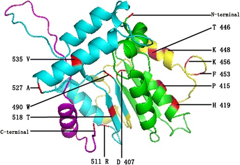
Figure 2.

Model building of the three-dimensional structure of the GRAS protein. The VHIID, LHRII, PFYRE, and SAW motifs are presented in green, yellow, blue, and pink, respectively. The figure was produced using the CPHmodels program, and amino acids refer to the AT3G54220 sequence.
