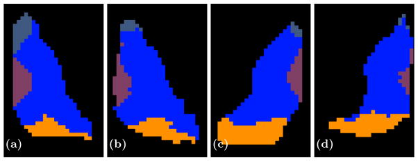
Fig. 4.

Shown are axial slices of (a) a manual delineation and (b) our parcellation for a right thalamus on one of our better results and (c) a manual delineation and (d) our parcellation of a left thalamus for a bad result. The AN is shown in a slate blue anterior to the thalamus, the VNG is the large blue body in the center of the thalamus, while MD and PUL are shown in purple and orange, respectively.
