
Fig. 3.
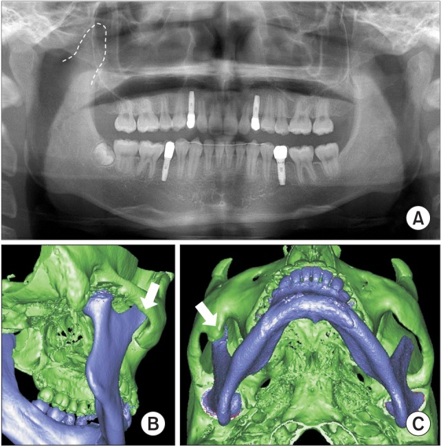
A. Preoperative panoramic radiograph showing hyperplasia of the right coronoid process. B, C. Preoperative computed tomography scans for case 2: three-dimensional view showing the elongated right coronoid process and heterotopic bone formation on the inside of the zygomatic bone (arrows).