Figure 1.

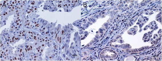
Simple tubular mammary carcinoma. Inmunohistochemical PR labelling is seen in the nuclei of tumour epithelial cells. A strong PR + tumour at day 1 (A) and a PR - tumour at day 15 (B) [3]. ABC immunohistochemical method. Bar = 10 μm.

Simple tubular mammary carcinoma. Inmunohistochemical PR labelling is seen in the nuclei of tumour epithelial cells. A strong PR + tumour at day 1 (A) and a PR - tumour at day 15 (B) [3]. ABC immunohistochemical method. Bar = 10 μm.