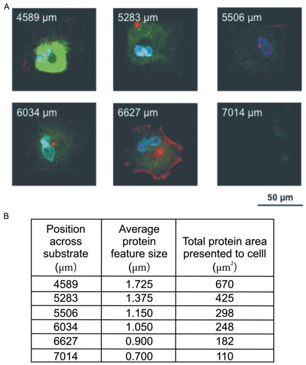
FIGURE 14.3.
Stem cell adhesion study using combinatorial fibronectin patterns to evaluate cell attachment and spreading. (A) A tilted polymer pen array (180 μm pen spacing) was used to generate 15 ×15 array of fibronectin features spaced by 4 μm. MSCs were cultured on these patterns for 1 week and subsequently stained for alkaline phosphatase (ALP) (green), actin (red), and the nucleus (blue). The labels on the resulting patterns shown in (B) indicate the x-position of patterns of a certain feature size across a substrate. The total area presented to the cell can be obtained by squaring the average protein feature size and multiplying by 225 for a 15 ×15 array of dot features.

