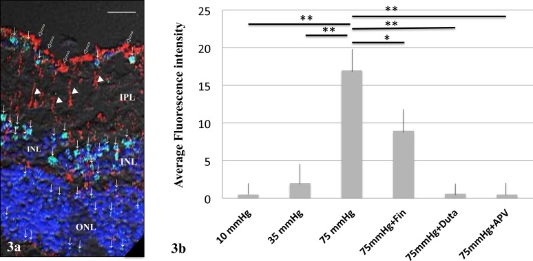
Figure 3.
(a) Immunofluorescent colocalization of allopregnanolone and vimentin by laser scanning microscopy. Allopregnanolone staining (green; FITC) was observed in the GCL, INL, and ONL at 75 mm Hg, and indicated by arrows. Antivimentin antibody (red; rhodamine) was localized to the Müller cell end feet (open arrows) and Müller cell bodies (white triangles). Double-labeled structures were not detected. Scale bars: 8 μm. (b) Summary of immunostaining studies shows fluorescence intensity by antiallopregnanolone antibody (arbitrary units) as mean ± SEM. Fluorescent intensity significantly increased at 75 mm Hg compared to 10 or 35 mm Hg. The increase in fluorescence induced by high pressure was significantly decreased by 1 μM finasteride (Fin). Administration of 1 μM dutasteride (Duta), almost completely inhibited the fluorescence increase. Administration of 50 μM APV also significantly decreased fluorescence. P values are calculated by unpaired Student's t-test compared to 75 mm Hg (*P < 0.001, **P < 0.0001).

