Figure 5.
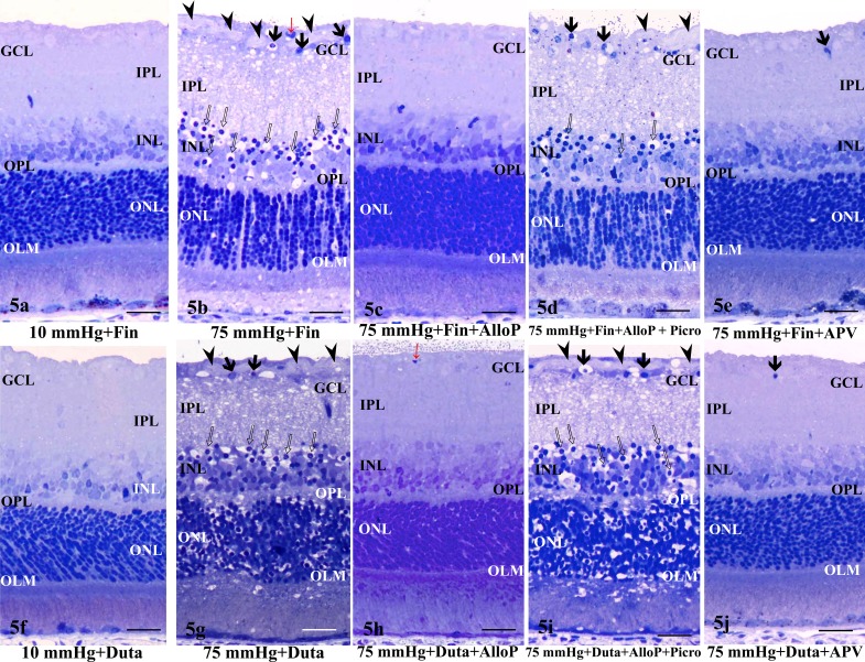
(a, b) Light micrographs of retinas incubated with 1 μM finasteride (Fin) at 10 (a) and 75 (b) mm Hg. (a) At 10 mm Hg, the retina incubated with finasteride showed no remarkable changes. (b) Administration with 1 μM finasteride induced excitotoxic changes characterized by bull's eye formation in the INL (open arrows) and edematous IPL along with axonal swelling in the NFL (arrowheads) in the retina at 75 mm Hg. Red arrow indicates blood capillary. Arrows indicate the degenerated ganglion cells. (c) Combination of 1 μM finasteride (Fin) and 1 μM allopregnanolone (AlloP) blocked the retinal excitotoxic degeneration at 75 mm Hg. (d) At 1 μM, picrotoxin (Picro) overcame the protective effect of 1 μM allopregnanolone in a pressure-loaded retina incubated with 1 μM finasteride at 75 mm Hg. Note the edematous IPL. Open arrows: Bull's eye formation in the INL. Arrowhead: Axonal swelling in the NFL. Arrows: Degenerated ganglion cells. (e) At 75 mm Hg, the excitotoxic changes induced by finasteride were inhibited in the retina incubated with 100 μM APV, but pyknotic ganglion cell nuclei remained (arrow). No remarkable changes were observed in other layers of the retina.
(f, g) Light micrographs of retinas incubated with 1 μM dutasteride at 10 (f) and 75 (g) mm Hg. (f) At 10 mm Hg, the retina incubated with dutasteride (Duta) showed no remarkable changes. (g) Administration with 1 μM dutasteride induced excitotoxic changes characterized by bull's eye formation in the INL (open arrows) and edematous IPL along with axonal swelling in the NFL (arrowheads) in the retina at 75 mm Hg. Arrows indicate the degenerated ganglion cells. (h) Combination with 1 μM dutasteride (Duta) and 1 μM allopregnanolone (AlloP) blocked the retinal excitotoxic degeneration at 75 mm Hg. Red arrow indicates blood capillary. (i) At 1 μM, picrotoxin (Picro) overcame the protection effect of 1 μM allopregnanolone in a pressure-loaded retina incubated with 1 μM dutasteride at 75 mm Hg. Open arrows: Bull's eye formation in the INL (red arrows). Arrowhead: Axonal swelling in the NFL. Arrow: Degenerated ganglion cells. (j) At 75 mm Hg, excitotoxic changes induced by dutasteride were inhibited in a retina incubated with 100 μM APV, but pyknotic ganglion cell nuclei remained (arrow). No remarkable changes were observed in other layers of the retina. (a–j) Scale bars: 15 μm.

