Figure 4.
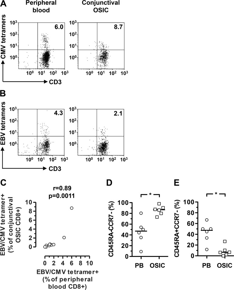
Frequency of cytomegalovirus (CMV)- and Epstein-Barr virus (EBV)-specific T cells among conjunctival epithelial CD8+ T cells reflects that of peripheral blood. Flow cytometry plots were gated on live CD45+CD56−CD8+ cells. Conjunctival CD8+ T cells were stained with pooled CMV or EBV peptide MHC class I tetramers. The inclusion of the CD3 marker provides confirmation that the gated cells are indeed T cells. Representative plots identifying CMV- and EBV-specific T cells are shown in (A, B), respectively. Correlation between frequencies of CMV- or EBV-specific CD8+ T cells in peripheral blood and conjunctiva is shown in (C). Differences between effector memory and effector memory RA subsets within the virus-specific T cell populations in peripheral blood and conjunctiva are shown in (D, E). Statistical analysis was undertaken by a Spearman correlation or Wilcoxon matched-pairs signed rank test. EBV, Epstein-Barr virus; CMV, cytomegalovirus; PB, peripheral blood; OSIC, conjunctival ocular surface impression cytology; *P < 0.05.

