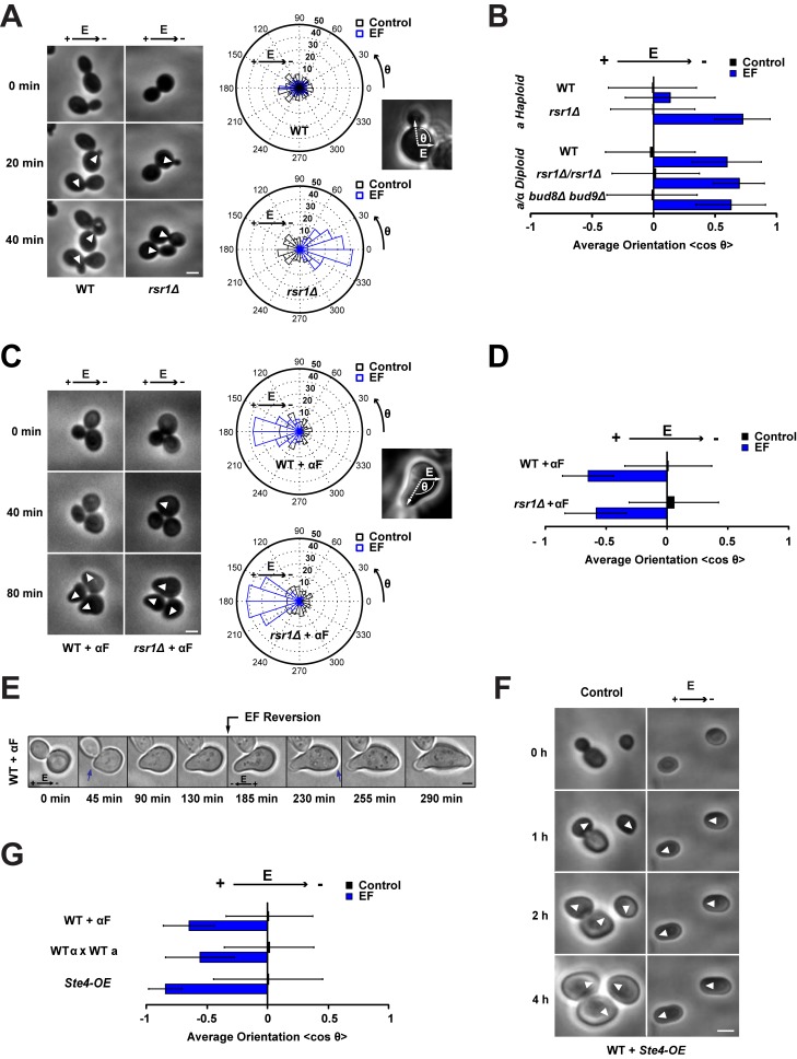
Figure 1. Budding versus shmooing yeast cells polarize in opposite directions in an electric field.
(A) Phase contrast time lapse of WT and rsr1Δ budding yeast cells growing under an EF of 50 V/cm. White arrowheads point at sites of bud emergence. On the right are radial histograms of polarized growth direction (indicated as the final angle of bud emergence with the EF, θ) for WT and rsr1Δ cells in the presence or in the absence of an EF. (B) Average bud orientation, computed as <cosθ> after 3 h of growth in the absence or in the presence of an EF, for a population of haploids and diploids of the indicated genotype. A positive average orientation represents an orientation to the cathode (negative electrode of the EF), whereas a negative orientation stands for an orientation to the anode. (C) Phase contrast time lapse of WT and rsr1Δ budding yeast cells growing mating projections (“shmoos”) in the presence of α-factor (αF) under an EF of 50 V/cm. White arrowheads point at sites of shmoo emergence. On the right are radial histograms of polarized growth direction. (D) Average shmoo orientation after 3 h of mating tip growth in the absence or in the presence of an EF for a population of WT and rsr1Δ cells treated with α-factor. (E) Time-lapse images of shmoo reorientation in a WT cell after inverting the EF direction. A second shmoo is formed at the new anodal side after reversing the EF. Blue arrows indicate sites of shmoo emergence. (F) Time lapse of WT cells overexpressing Ste4p (Ste4-OE) in the absence or in the presence of an EF of 50 V/cm. White arrowheads point at sites of polarized growth. (G) Average shmoo orientation after 3 h in the absence or in the presence of an EF for a population of WT cells treated with α-factor, WT mating pairs, and WT cells overexpressing Ste4p. n>50 cells for all conditions. Error bars represent standard deviations. Scale bars: 2 µm.

