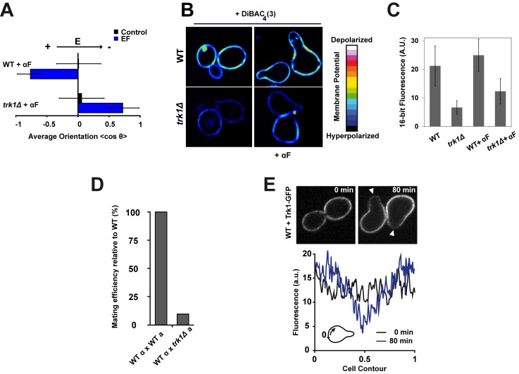
Figure 3. A potassium transporter, Trk1p, mediates EF response in shmoos.
(A) Average shmoo orientation after 3 h in the absence or in the presence of an EF for a population of WT and trk1Δ cells treated with α-factor (αF) (n>50 cells). (B) Sixteen-color images of WT and trk1Δ cells stained with the membrane-potential-sensitive dye DiBAC4(3), which depicts reduced membrane fluorescence upon membrane hyperpolarization. (C) Quantification of DiBAC4(3) dye membrane staining intensity in WT and trk1Δ cells. (D) Mating efficiency of trk1Δ cells relative to WT. (E) Confocal single focal plane time-lapse images of Trk1-GFP in WT cells grown in the presence of α-factor. White arrowheads indicate shmoo growth sites. Below is the mean fluorescence intensity along the cell contour at times 0 and 80 min after α-factor treatment, averaged on five independent cells. Distances are normalized between 0 and 1 so that the value 0.5 corresponds to the site of shmoo emergence.

