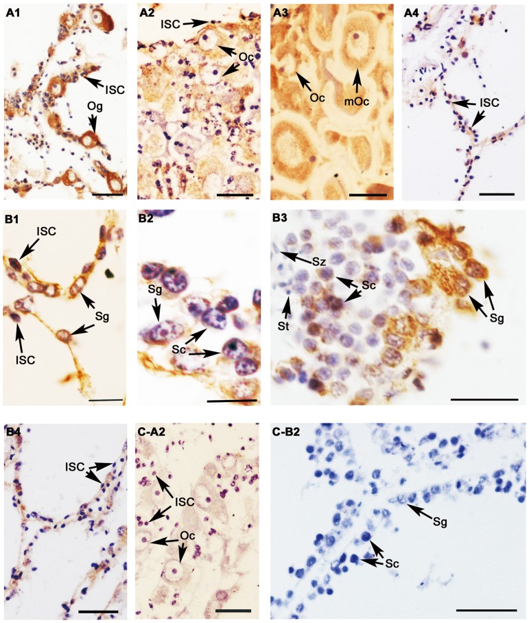
Figure 5. Location of β-catenin protein in C. farreri gonads detected by immunohistochemistry.
A, B: Positive signals are brown and represent anti-β-catenin in ovaries (A) and testes (B). C: Negative control with preimmune serum; 1: proliferative stage; 2: growth stage; 3: mature stage; 4: resting stage; ISC: intragonadal somatic cell; Og: oogonium; Oc: oocyte; mOc: mature oocyte; Sg: spermatogonium; Sc: spermatocyte; St: spermatid; Sz: spermatozoon; Scale bars: B1, B2, and B3 are 10 µm, others are 30 µm.

