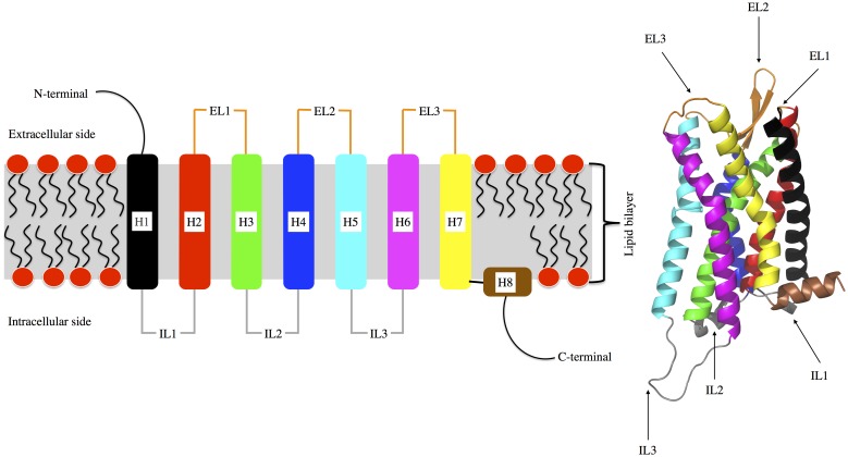
Figure 1. Left: Schematic view of the overall structure of a G protein-coupled receptor (GPCR), with depiction of the connectivity of the intracellular (IL) and extracellular (EL) loops between helices (H).
Right: 3D structure of the reconstructed µOR structure (see “Methods”). Color codes of ILs, ELs, and Hs are identical in both left and right pictures.

