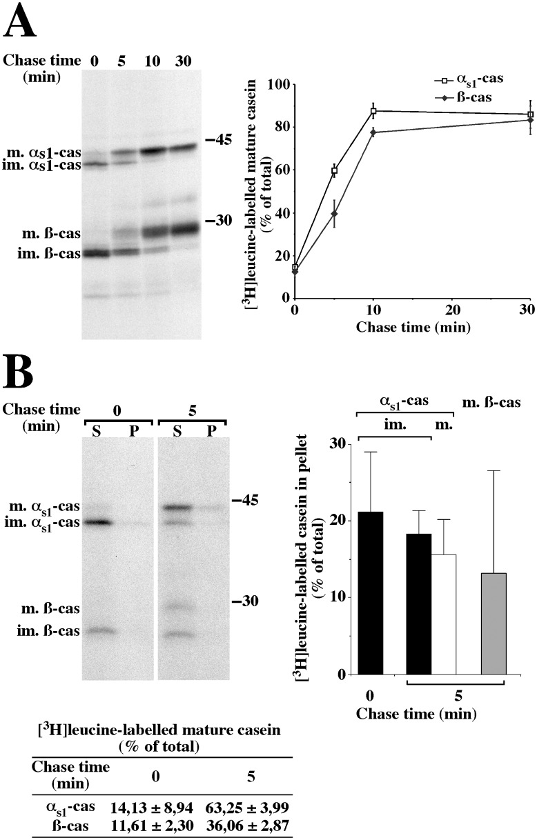
Figure 1. A membrane-associated form of αs1-casein is also present in the Golgi apparatus of rat MECs.
(A) Time course for the arrival of newly synthesised caseins in the Golgi apparatus. Rat mammary gland fragments were pulse-labelled for 3 minutes with [3H]leucine and chased for the indicated times. At the end of the various chase periods, a PNS was prepared from the cells and analysed via SDS-PAGE and fluorography, followed by quantification of the immature (im.) and mature (m.) forms of both αs1- and ß-casein (cas). The amount of the mature form of the caseins was expressed as percent of total (sum of immature and mature forms). The mean ± s.d. from three independent experiments is shown. (B) Relative proportions of membrane-associated forms of the caseins in the ER and the Golgi apparatus. Rat mammary gland fragments were either pulse-labelled for 3 minutes with [3H]leucine or pulse-labelled and chased for 5 minutes. Aliquots of the PNS prepared from the cells were subjected to centrifugation and the resulting membrane pellet was resuspended and incubated for 30 minutes in non-conservative buffer in the presence of saponin. After centrifugation, supernatants (S) and pellets (P) were analysed via SDS-PAGE and fluorography, followed by quantification of the immature (im.) and mature (m.) forms of both αs1- and ß-casein. The amount of the mature form of the caseins (Table in panel B) was expressed as percent of total (sum of immature and mature forms). The amount of the various forms of the caseins in pellet (bar graph) is expressed as percent of total (sum of pellet and supernatant). The mean ± s.d. from three independent experiments is shown. Representative fluorograms are shown. Relative molecular masses (kDa) are indicated.

