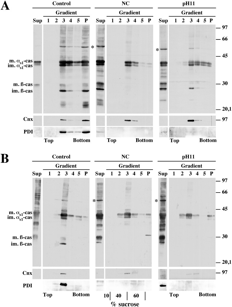
Figure 5. Purification of membrane-associated-αs1-casein fraction from rat mammary gland tissue on sucrose step gradients.
A purified rough microsome fraction (A) or membrane-bound organelles from a PNS (B), both prepared from rat mammary gland tissue, were incubated in the absence (Control) or in the presence of saponin under non-conservative conditions (NC) or under carbonate buffer at pH 11.2 (pH 11). After centrifugation, supernatants were collected and membrane pellets were subjected to flotation on a sucrose step gradient (theoretical sucrose concentrations are indicated at the bottom of the central gel in panel B). Half of the supernatant (Sup), gradient fractions collected from the top (1 to 5) and gradient pellet (P) were analysed via SDS-PAGE followed by immunoblotting with polyclonal antibodies against either mouse milk proteins. Representative ECL signals from 5 (microsomes) or 3 (PNS) independent organelle preparations are shown. The distribution of Cnx and PDI was analysed within the above immunoblots. Relative molecular masses (kDa) are indicated. im. αs1-cas: immature αs1-casein; m. αs1-cas: mature αs1-casein; im. ß-cas: immature ß-casein; m. ß-cas: mature ß-casein; *: protein band with electrophoretic mobility identical to PDI.

