
FIG. 9.
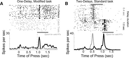
Stimulus-related activity in dmPFC.A: peristimulus rasters and histograms from a neuron with stimulus-related firing in dmPFC. Activity is shown for trials with a stimulus presented at a delay of 1.0 s (black) and for time production (or “catch”) trials (gray), on which the stimulus was not presented and rats were rewarded if they held the lever down for ≥1.0 s. Approximately 14% of neurons in dmPFC showed this pattern of firing and no neurons like this were found in motor cortex. B: activity from a dmPFC neuron in two-delay sessions that fired in response to the stimulus, similar to the neuron in A (0.4-s delays: gray; 1.0-s delays: black). This neuron also fired on some trials with long delays at the time of the short delay (black arrow) and these spikes are suggestive of stimulus-anticipatory activity. Note that all analyses are restricted to correct trials with a long delay.