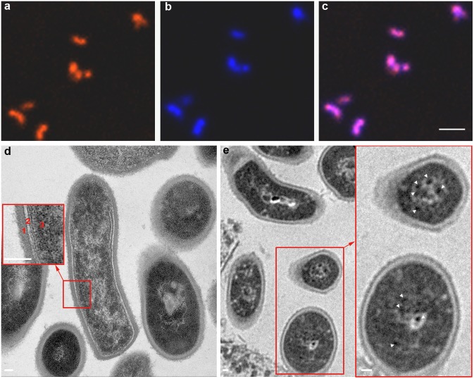
Figure 2. Representative confocal fluorescence microscopy and TEM images of Texas Red iron-oxide labeled C.novyi-NT.
Texas Red particles were observed within the C.novyi-NT (a). C.novyi-NT were stained blue with DAPI (b). Merged image (c) demonstrates the co-registration of Texas Red and DAPI staining. TEM of control unlabeled C.novyi-NT (d) with inset depicting the bacterial wall (1), light plasma membrane (2), and relatively homogeneous dark-stained cytoplasm and nucleoid core (3). Within TEM image of iron-oxide labeled C.novyi-NT (e), a large number of punctate dark-stained iron granules were observed within the bacteria (white arrowheads within inset). Scale bars: a, b, and c = 6 µm; d, e, and inset in d and e = 60 µm.

