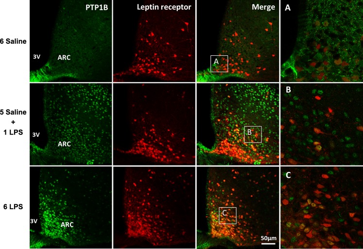
Fig. 2.
Representative photomicrographs showing PTP1B expression (green) in the hypothalamic arcuate (ARC) nucleus 2 h after the last injection in LepRb reporter mice (red) injected once a day with 6 doses of saline, single dose of LPS (100 μg/kg ip), or repeated doses of LPS. 3V, third ventricle. Scale bar, 50 μm. Insets (A, B, and C): examples of LepRb neurons coexpressing or not PTP1B, ×40 magnification.

