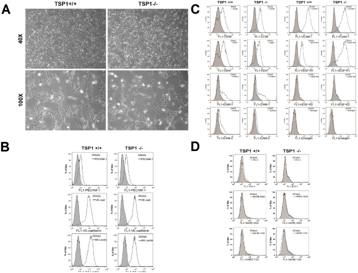
Figure 1. Isolation and characterization of mouse choroidal endothelial cells (ChEC).
Thrombospondin1 (TSP1)+/+ and TSP1−/− ChEC were prepared as described in MATERIALS AND METHODS and cultured on gelatin-coated plates in 60-mm dishes. A: cells were photographed in digital format at ×40 and ×100 magnification. Note TSP1−/− ChEC exhibited a similar elongated and spindly morphology compared with TSP1+/+ ChEC. B: The expression of vascular EC markers in ChEC. ChEC were examined for expression of PECAM-1, VE-cadherin (VE-cad), and B4 lectin by FACS analysis. Shaded areas show control IgG staining. Note the similar expression of these cellular markers in both cells. C: FACS analysis for expression of other cell surface markers. Please note expression of CD36, CD 47, ICAM-1, ICAM-2, and VCAM-1 expression in these cells. We also detected significant expression of VEGF-R1 in these cells whose level was increased in TSP1−/− ChEC. The VEGF-R2 expression was almost undetectable. D: FACS analysis of EC markers for fenestration, PV-1 and HTAR (stabilin-2). Please note minimal expression of these markers. These experiments were repeated at least twice with two different isolations of choroidal EC, with similar results.

