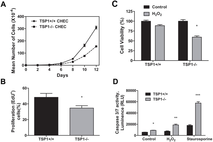
Figure 3. Altered proliferation and apoptosis of TSP1−/− ChEC.
A and B: the rate of ChEC proliferation was determined by counting the number of cells in triplicate after different days in culture as described in Methods (A) and by analyzing the rate of DNA synthesis by FACScan flow cytometry analysis (B; P<0.05; n = 3). C: Hydrogen peroxide (H2O2) toxicity of ChEC was measured by MTS assay. ChEC were incubated with 1 mM H2O2 in EC growth medium for 2 days in 96-well plates and subjected to the MTS assay. TSP1−/− ChEC were significantly more sensitive to cytotoxic effect of H2O2 (*P<0.05; n = 3). D: The rate of apoptosis was determined by measuring caspase activity with luminescent signal from caspase-3/7 DEVD-aminoluciferin substrate, as recommended by the supplier. As an apoptotic stimulus, H2O2 and staurosporine in EC growth medium were added for 8 h. Please note the significant increase in the rate of apoptosis in TSP1−/− ChEC compared with TSP1+/+ cells (*,**,*** P<0.05; n = 3). RLU, Relative Light Unit.

