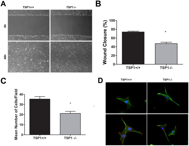
Figure 4. TSP1−/− ChEC are less migratory.
A: Cell migration was determined by scratch wound assay of the ChEC monolayers on gelatin-coated plates. Wound closure was monitored by photography within 48 h. B: Quantitative assessment of the data (*P<0.05, n = 3). C: Cell migration was also determined using a transwell migration assay (*P<0.05, n = 3). D: The indirect immunofluorescence staining of phalloidin (green; actin) and vinculin (red; focal adhesions). Please note similar actin stress fibers and focal adhesion organizations in TSP1+/+ and TSP1−/− ChEC. The quantitative assessment of fluorescence intensities showed no significant differences (P>0.05; n = 3; not shown). These experiments were repeated with two different isolations of cells with similar results.

