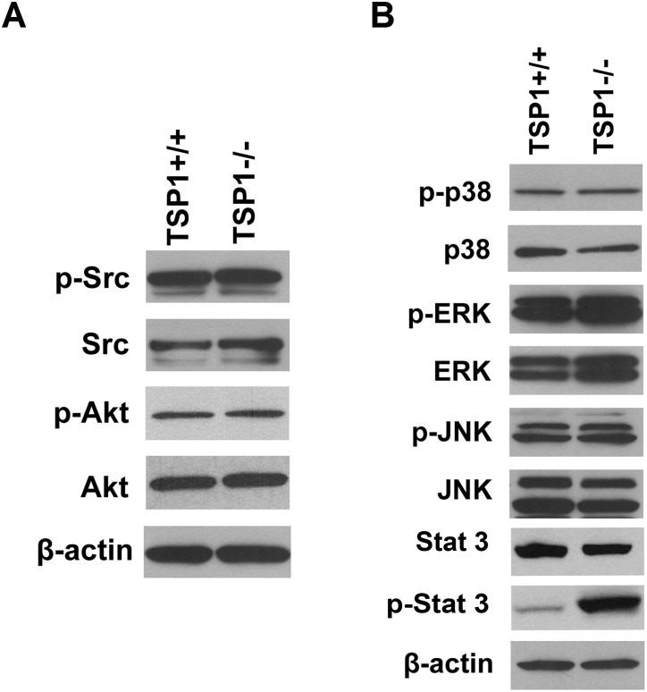
Figure 11. Expression and phosphorylation of Src, Akt, and MAPKs signaling pathways in ChEC.
Expression and phosphorylation of Src and Akt were analyzed by Western blotting (A). A similar level of phosphorylated and total Src and Akt was observed in TSP1+/+ and TSP1−/− choroidal EC. Expression and phosphorylation of ERKs, JNK and p38 MAP kinases were analyzed by Western blotting (B). Please note minimal impact of TSP1-deficincy on phosphorylation and expression of ERKs in ChEC. A significant increase in phosphorylation of STAT3 was observed in TSP1−/− ChEC, while total level of STAT3 was not affected. These experiments were repeated with two different isolations of cells with similar results.

