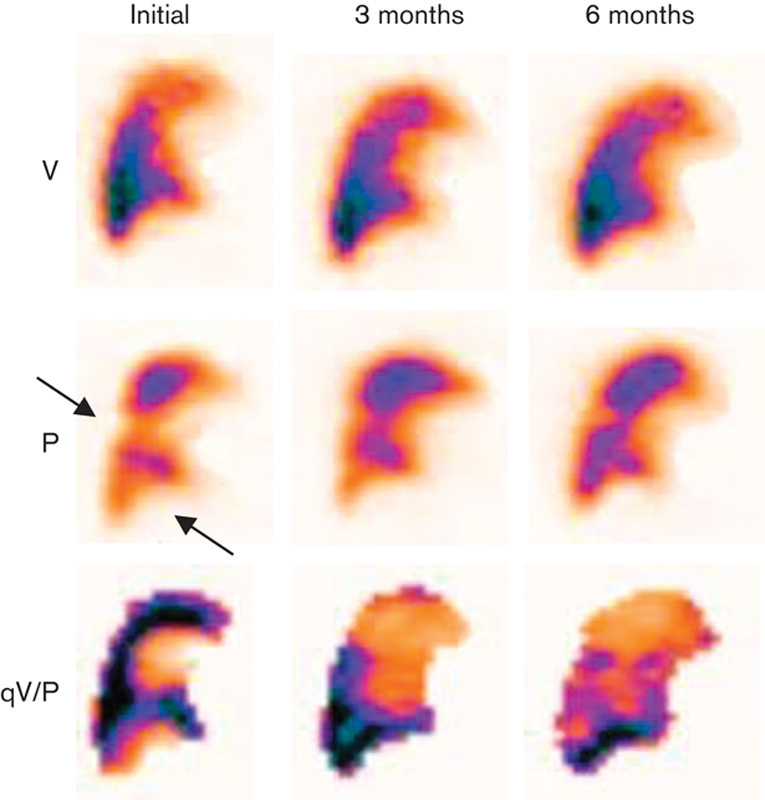
Fig. 3.

Sagittal V/P SPECT images of a patient with PE (arrow) at initial examination. Only partial regression of the perfusion defects is seen during the follow-up period and there remain perfusion defects at 6 months. P, perfusion; PE, pulmonary embolism; qV/P, V/P quotient; V, ventilation; V/P SPECT, ventilation/perfusion single-photon emission computed tomography.
