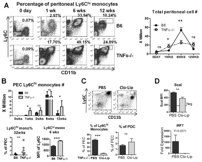
Figure 3. TNFα affects Ly6Chi (inflammatory) monocytes.
A, Peritoneal cells were analyzed by flow cytometry 0–12 weeks after pristane injection. Left: Percentages of CD11b+Ly6Chi monocytes (rectangles) in total peritoneal exudate cells were assessed by flow cytometry. Right, total peritoneal cell counts in TNFα−/− (open circles) and B6 (closed circles) mice at 0–12 weeks after pristane treatment. B, Top: Comparison of CD11b+Ly6Chi monocytes at different time-points (3–5 mice per group). Bottom left, Ly6Chi monocytes (CD11b+Ly6ChiLy6G−) as a percentage of total peritoneal exudate cells at 32 weeks. Bottom right, Ly6C expression (MFI) on peritoneal Ly6Chi monocytes. C, Top: Flow cytometry of pristane-elicited peritoneal cells 2-days following treatment with clodronate liposomes (Clo-Lip) or PBS. Boxed areas indicate the percentage of Ly6Chigh monocytes (4 mice per group). Bottom left and right: Percentage of peritoneal Ly6Chi monocytes (left) and pDCs (right) in PBS or Clo-Lip treated mice. D: MFI of ScaI expression (flow cytometry) on peritoneal B cells and expression of IRF7 mRNA (Q-PCR) after PBS or Clo-Lip treatment in peritoneal exudate cells. * P < 0.05; ** P < 0.01; *** P < 0.001 unpaired Student’s t test.

