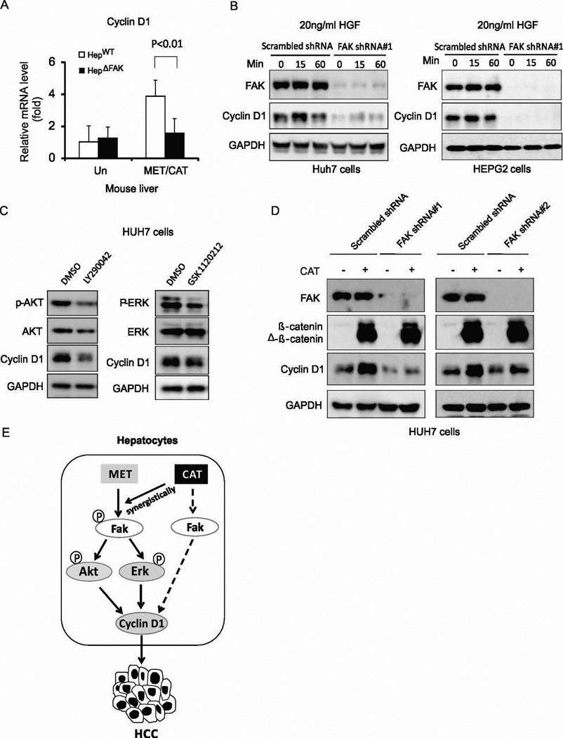
Figure 7. FAK mediates Cyclin D1 regulation by MET or CAT in HCC cells.
(A) Cyclin D1 mRNA expression level in the livers of HepWT and HepΔFAK mice 7 weeks after injection of MET/CAT. (B) Protein expression of FAK, Cyclin D1 and GAPDH in Huh7 and HEPG2 cells infected with scrambled shRNA or FAK shRNA#1 lentiviral particles, then treated with 20ng/ml HGF for 15 or 60 minutes. (C) Protein expression of p-AKT, AKT, p-ERK, ERK, Cyclin D1 and GAPDH in Huh7 cells treated with 10 µM LY294002 or 500 nM GSK1120212 for 24 hours. (D) Protein expression of FAK, Cyclin D1 and GAPDH in Huh7 cells infected with scrambled shRNA, FAK shRNA#1 or FAK shRNA#2 lentiviral particles, then transfected with CAT or control plasmids. (E) A schematic model: hydrodynamic injection of MET into hepatocytes results in Fak activation. CAT does not activate FAK directly but the combination of CAT and MET has a synergistic effect on Fak activation. Fak activation regulates MET- or CAT-mediated Cyclin D1 induction, acting through either Akt- and Erk-dependent or -independent pathways, thereby promoting hepatocarcinogenesis.

