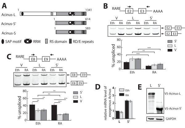
Figure 1. Both Acinus-S’ and Acinus-L cooperate with RA to activate the splicing of a RA-responsive reporter minigene containing a weak 5′ splice site.
A. Functional domains of the three human Acinus isoforms. B. Acinus does not facilitate the splicing of RARE-tuba1bE2-E3 pre-mRNA containing a strong 5′splice site. C. Acinus and RA cooperatively activate the splicing of RARE-tubg1E8-E9 pre-RNA containing a weak 5′splice site. D. Relative level of exogenous Acinus-L and Acinus-S’ mRNA in transfected cells. E. Relative level of exogenous Acinus-L and Acinus-S’ in transfected cells. 293A cells were transfected with pRARE-tuba1bE2-E3 (B) or pRARE-tubg1E8-E9 (C-E) along with the expression vector DNA for RARβ (B–E) and the expression vector DNA for either V5-Acinus-L or V5-Acinus-S’ (B-E). The V5 empty vector DNA (pcDNA3.1nV5DEST) was transfected as a control for pV5-Acinus-L or pV5-Acinus-S’ (B-E). In addition, the internal control Renilla vector DNA (pRL-CMV) was transfected as a normalizer for transfection efficiency (B-E). Twenty-four hr following transfection, cells were treated with ethanol (B-E) or RA (10−6 M) (B-D) for 24 hr. RNA was prepared and used to analyze splicing in Panels B-C or relative mRNA levels in Panel D. In Panels B-C, RT-PCR was performed to analyze splicing of the minigene pre-mRNAs using the indicated forward and reverse primers (see diagram for location). Spliced and unspliced PCR products were resolved on polyacrylamide gels, stained using SYTO 60 and quantitated using LI-COR Odyssey Infrared Imaging System. Percentage of unspliced was calculated as the ratio of the intensity of PCR products of the unspliced transcripts to that of the sum of the spliced and unspliced transcripts. In Panel D, RT-qPCR was performed to quantitate the relative mRNA levels of exogenous Acinus-L and Acinus-S’. The mRNA levels were normalized to Renilla mRNA levels. The normalized relative mRNA level of exogenous Acinus-L coexpressed with RARE-tubg1E8-E9 in cells treated with ethanol was set to 1. The forward primer is located within the C-terminal common region of Acinus isoforms and the reverse primer is located within the bovine growth hormone polyadenylation region on the V5 vector. In Panel E, whole protein extracts were isolated and western blot was performed using anti-V5 or anti-GAPDH antibodies. V, V5 empty vector; L, V5-Acinus L; S’ V5-Acinus-S’; Eth, ethanol; RA, retinoic acid. Values represent mean ± SD from 3 independent experiments. ** p < 0.01, *** p < 0.001, unpaired t test.

