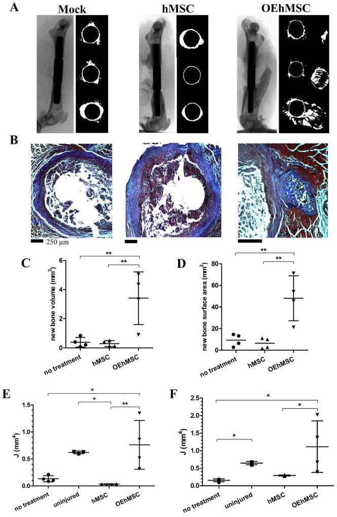
Fig. 2. A comparison of directly administered hMSCs and OEhMSCs for healing a critical-sized segmental defect of the femur and allowed to heal for 2 weeks.
MSCs were directly administered to pin-stabilized defects in the presence of clotting plasma only. Panel A: μCT scans (left) and axial reconstructions of the proximal edge of the defect, (top right), mid-defect (center right) and distal edge of the defect (bottom right). Panel B: paraffin embedded axial sections stained with Masson’s trichrome corresponding to the mid-defect. Panel C and D: measurements of new bone volume (panel C) and new bone surface area (panel D) between the original edges of the defect calculated by μCT-rendered reconstructions. Panel E: measurements of the polar moment of inertia (J) at the mid-defect (values are generated by taking the mean of 3 adjacent axial images). Panel F: the mean value of J for the entire ROI. Statistical testing was performed by ANOVA and Tukey post-test. P-values key: P<0.05 = *, P<0.01=**, P<0.005=***.

