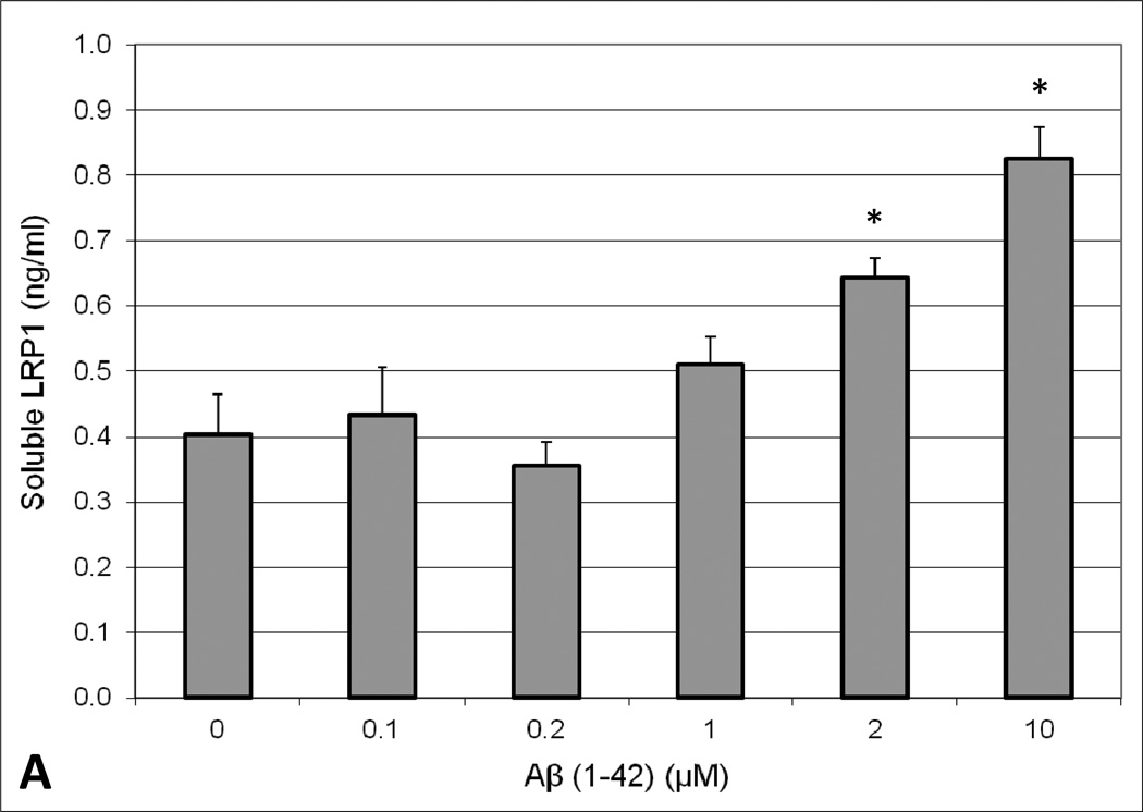
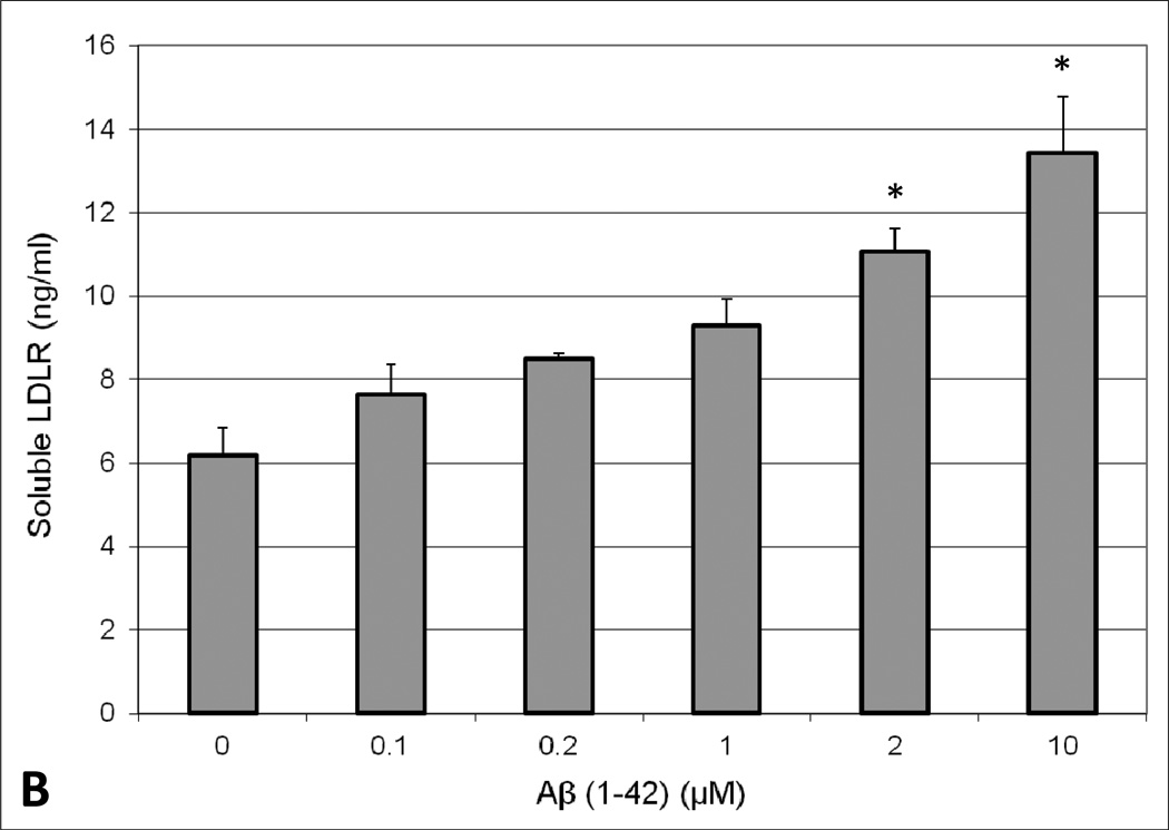
Fig. 1.
Appearance of extracellular soluble (A) LRP1 or (B) LDLR in human brain endothelial cells (HBMEC) upon treatment with Aβ (1–42). HBMEC were exposed to various concentrations (0.1, 0.2, 1, 2, and 10 µM) of human Aβ(1–42) for 48 hours at 37°C. Following the treatment period, the extracellular media was collected and analyzed for LRP1 or LDLR content by ELISA. Values represent mean ± SEM (n = 3) and are expressed as ng of LRP1 or LDLR per ml of media. *P < 0.05 compared to control as determined by ANOVA and Bonferroni post-hoc test.


