Fig. 3.
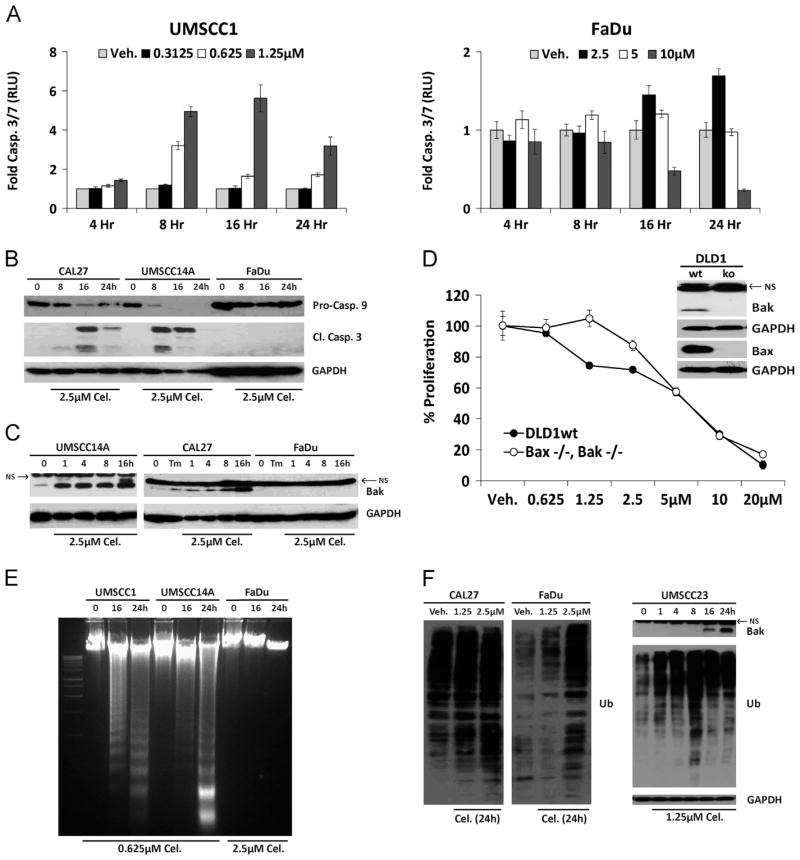
Celastrol induces apoptosis in OSCC cells. A. Luminescent caspase 3/7 assays performed with celastrol-sensitive (UMSCC1) and celastrol-resistant (FaDu) cells; error bars represent standard deviation of biological replicates. B. Immunoblot analysis of whole-cell lysates from celastrol-treated OSCC cells with polyclonal antibodies for Caspases-9 and -3. C. Immunoblot analysis of whole-cell lysates from OSCC cells with polyclonal antibody for Bak; NS=non-specific band present in all OSCC and DLD1 cells, including Bak−/−. D. Proliferation of wildtype or zinc finger deleted DLD1 Bax−/−/Bak−/− colorectal adenocarcinoma cells treated for 24 h (Two-way ANOVA, P value <0.0001 for dose and interaction). E. Electrophoretic resolution of genomic DNA harvested from OSCC cultures to appreciate DNA laddering following celastrol exposure. F. Immunoblot analysis of ubiquitinated (total) proteins in whole cell lysates after 24 h (left); accumulation of ubiquitinated proteins in UMSCC23 cells preceded Bak accumulation (right). For Immunoblot analysis, each membrane was stripped and re-probed with monoclonal GAPDH.

