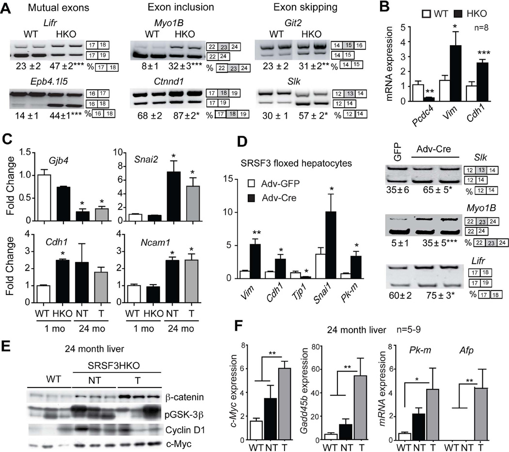
Fig. 6. Loss of SRSF3 leads to aberrant splicing of EMT genes, activation of β-catenin signaling and c-MYC expression, and induction of liver progenitor cell markers.
(A) Analysis of splicing of EMT-associated transcripts (Lifr, Epb4.1l5, Myo1b, Ctnnd1, Git2 and Slk) at 1 mo. Representative gels are shown with the percentage of exon use given below the gels. The exons involved in the alternative splicing event are shown to the right of the gel images. (B) Quantification of mRNA expression of the EMT-associated genes programmed cell death 4 (Pcdc4), vimentin (Vim) and E-cadherin (Cdh1) at 1 mo by Q-PCR. (C) Quantification of gap-junction protein 4 (Gjb4), slug/snail2 (Snai2), Cdh1 and neuronal cell adhesion molecule 1 (Ncam1) by microarray. Results are presented as mean fold change vs. WT ± SEM. (D) Change of EMT gene expression and alternative splicing following Adv-cre infection of primary Srsf3:floxed hepatocytes. (E) Immunoblotting for non-phospho β-catenin, phospho-GSK3β, Cyclin D1 and c-Myc in livers at 24 months. Three representative samples per group are shown. (F) Quantification of c-Myc, Gadd45, Pkm and Afp mRNA expression at 24 mo by Q-PCR. T and NT indicate tumor and non-tumorous areas of HKO liver.

