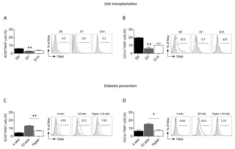
Figure 2.
TIM4 expression on APCs was evaluated by flow cytometry during the allo- (A, B) and autoimmune (C, D) anti-islet response in vivo. A decrease in the percentage of B-cells positive for TIM4 (n=3, **p<0.01 vs. D0; A) and DCs positive for TIM4 (n=3, **p<0.01 vs. D0; B) was evident after fully-mismatched islet transplantation of BALB/c islets into C57BL/6 recipients. In diabetes prevention studies, a reduction in the percentage of B-cells (n=3, **p<0.01 vs. 10wks; C) and DCs (n=3, *p<0.05 vs. 10wks; D) positive for TIM4 was observed in hyperglycemic mice.

