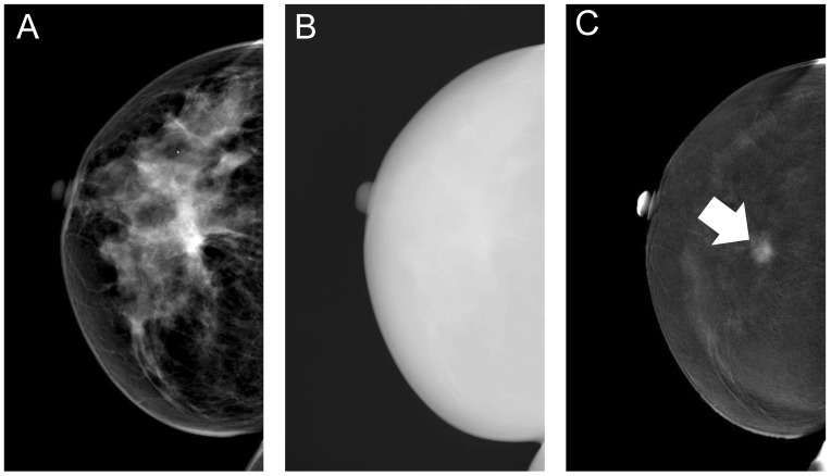
Figure 1.
Typical example of the different contrast-enhanced spectral mammography images. First, a low-energy image is acquired (A), immediately followed by the high-energy image (B), which is used in post-processing to create the recombined image (C), in which the invasive breast cancer is clearly visible (arrow).

