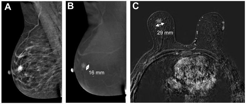
Figure 5.
Example of poor agreement between CESM and breast MRI. The low-energy CESM image (A) shows an ill-defined mass behind the nipple, enhancing on the recombined images (B, 16 mm). Breast MRI showed a spiculated mass (C, 29 mm). Histopathological results showed a 21 mm invasive lobular carcinoma.

