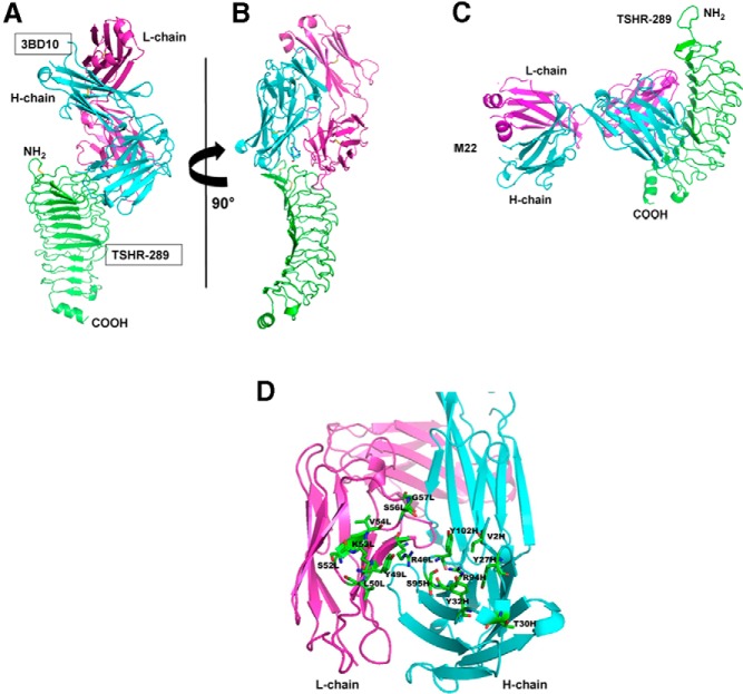
Figure 4.
A, Docking of the 3-dimensional crystal structures of the 3BD10 Fab and TSHR-289. The latter represented TSHR amino acid residues 22–260 (2XWT) (16) extended to TSHR residue 289 by modeling based on the homologous region of the FSHR (30). Cyan, 3BD10 H-chain; magenta, 3BD10 L-chain; green, TSHR-289. B, Rotation of molecules by 90°. C, Human monoclonal TSAb Fab M22 docked to TSHR-289 indicates no overlap with the 3BD10 epitope. Docking by Z-dock used the 3-dimensional structure of M22 (3GO4) (15) and TSHR-289. Docking was guided by including all the contact residues between M22 and TSHR-260 (15). D, 3BD10 residues at the binding surface to TSHR-289.

