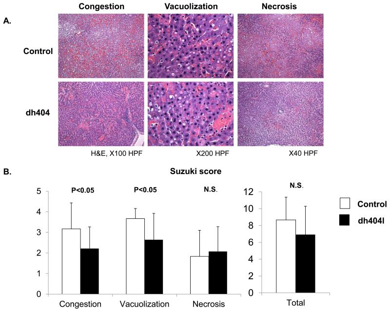
Figure 1. Liver damage evaluation by histological assay.
(A). Liver tissue specimens were stained with hematoxylin and eosin. Representative specimens are shown (×40 - ×200). (B) Suzuki score The level of sinusoidal congestion and vacuolization in the dh404 treated group was significantly improved compared to the untreated group but not necrosis level. There was no significant difference in the total score. Data represent mean±S.D.

