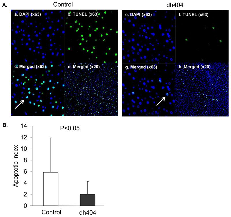
Figure 2. TUNEL staining in liver post ischemia/reperfusion injury.
(A). a-d. TUNEL staining in the control group (a; DAPI ×63, b; TUNEL×63, c; Merged×63, d; Merged ×20). e-h. Dh404 group. (Arrows indicate apoptotic cells) Apoptotic index indicates that the dh404 treated group had significantly less TUNEL-positive cells when compared to the control group (P<0.05). (B). Apoptotic index. Data represent mean±S.D.

