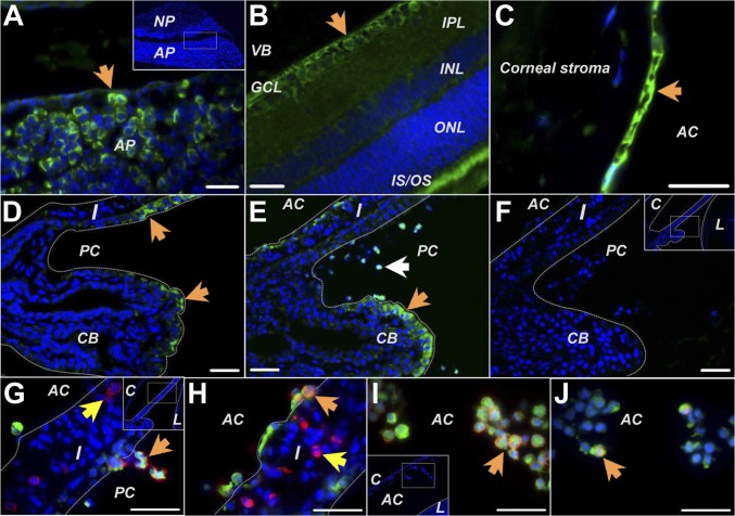
Fig. 3.
Immunohistochemical studies of GHRH-R expression in pituitary and ocular tissues. (A) GHRH-R was found on cells (arrow) in the anterior pituitary gland. (B) In the retina, GHRH-R was stained in the ganglion cell layer (arrow) and outer and inner segments of photoreceptors. (C) GHRH-R was observed on the corneal endothelium (arrow). (D) In normal rats GHRH-R was expressed on epithelial cells in the ciliary body process and iris. (E) In LPS-treated animals GHRH-R expression was up-regulated on cells in the ciliary epithelium (orange arrow) and on cells infiltrating into the anterior and posterior chambers (white arrow). (F) Treatment without the primary antibody gave no staining on the LPS-treated eye sections. (G–J) GHRH-R (green) was intensely stained on the cells expressing the leukocyte marker CD43 (red in G and I) or the monocyte/macrophage marker CD68 (red in H and J) when they detached from the iris (orange arrows in G and H) or resided in the aqueous humor (arrows in I and J). GHRH-R was not found in the stroma cells of the iris (yellow arrows in G and H). AC, anterior chamber; AP, anterior pituitary gland; C, cornea; CB, ciliary body; GCL, ganglion cell layer; I, iris; PC, posterior chamber; IPL, inner plexiform layer; IS/OS, inner and outer segment of photoreceptors; INL, inner nuclear layer; L, lens; NP, neurohypophysis; ONL, outer nuclear layer; VB, vitreous body. (Scale bars: 35 µm.)

