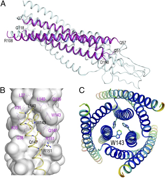
Fig. 4.
Crystallographic structure of the covNHR3–ABC construct. (A) Cartoon representation of the superposition of covNHR3–ABC (purple) onto the theoretical model of the gp41 ectodomain in the postfusion conformation (PDB ID code 1IF3; gray) (21) and the three NHR helices of 5-Helix (PDB ID code 2XRA; blue) (7). (B) Upper view of the packing of three covNHR3–ABC molecules in the crystal. The side chains of W143 form an aromatic cluster that brings together three covNHR3–ABC molecules. The helices are colored by B-factor spectrum (from blue to red). (C) Conserved hydrophobic pocket on the surface of the two parallel helices of covNHR3–ABC. The side chains of residues lining the pocket are labeled in magenta. The helix of another covNHR3–ABC molecule (in yellow) packs in the crystal against this hydrophobic cavity with the insertion the side chain of W143. All of the figures were made using Pymol.

